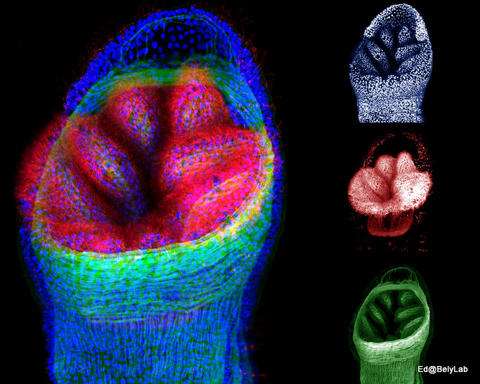
Dero digitata - Z-stack confocal projection of branchial pavillion

Description:
This image shows a three-channel confocal laser microscope image of the posterior end of a Dero digitata. Muscle actin is shown in green, cilliary tubulin is shown in red, and DNA is shown in blue. To the left side is the composite of all three channels; the individual channels are on the right.
Creator:
Zattara, Eduardo
Taxonomic name:
